
Description
The Integumentary System, by Bryan E. Anderson, MD, takes a concise and highly visual approach to illustrate the basic sciences and clinical pathology of the skin, hair and nails. This newly added, never-before-published volume in The Netter Collection of Medical Illustrations (formerly the CIBA "Green Books") captures current clinical perspectives on the integumentary system - from normal anatomy and histology to pathology, dermatology, and common issues in plastic surgery and wound healing. Using classic Netter illustrations and new illustrations created in the Netter tradition, as well as a great many cutting-edge histologic micrographs and diagnostic images, it provides a vivid, illuminating, and clinically indispensable view of this body system.- Gain a rich, holistic clinical view of every structure by seeing classic Netter anatomic illustrations, cutting-edge histologic images and diagnostic imaging studies side by side.- Visualize the most recent topics in cutaneous pathology such as sporothrix and cutaneous t-cell lymphoma as well as classic problems like alopecia and neurofibromatosis, informed by the latest developments in molecular biology and histologic imaging.- See current dermatologic concepts captured in the visually rich Netter artistic tradition via major new contributions from Netter disciple Carlos Machado, MD - making complex concepts easy to understand and remember through the precision, clarity, detail, and realism for which Netter's work has always been known.- Get complete, integrated visual guidance on the skin, hair, and nails in a single source, from basic sciences and normal anatomy and function through pathologic conditions.- Adeptly navigate current controversies and timely topics in clinical medicine with guidance from the Editor and informed by an experienced international advisory board.
Table of Contents
Section 1 - Anatomy, Physiology, and Embryology- Embryology of the skin- Normal Skin Anatomy- Normal Skin Histology- Skin Physiology - The Process of Keritinization- Normal skin flora- Vitamin D metabolism- Photobiology- Wound Healing- Morphology: Lichen Simplex Chronicus, Urticaria, and Postauricular Fissures- Morphology: Vitiligo, Tinea Faciei, and HerpesSection 2 - Benign GrowthS- Acrochordon- Becker's Nevus (smooth muscle hamartoma)- Dermatofibroma (sclerosing hemangioma)- Eccrine Poroma- Eccrine Spiradenoma- Eccrine Syringoma- Ephelide and Lentigines- Ephelide and Lentigines (Continued)- Epidermal Inclusion Cyst- Epidermal Nevus- Fibrofolliculoma- Fibrous Papule- Ganglion Cyst- Glomus Tumor and Glomangioma- Hidradenoma Papilliferum- Hidrocystoma- Keloid and Hypertrophic Scar- Leiomyoma- Lichenoid Keratosis- Lipoma- Median Raphe Cyst- Melanocytic Nevi: Blue Nevi- Melanocytic Nevi: Common Acquired Nevi and Giant Congenital Melanocytic Nevi- Melanocytic Nevi: Congenital Nevi- Milia- Neurofibroma- Nevus Lipomatosus Superficialis- Nevus of Ota and Nevus of Ito- Nevus Sebaceus- Osteoma Cutis- Palisaded Encapsulated Neuroma- Pilar Cyst (Trichilemmal Cyst)- Porokeratosis- Pyogenic Granuloma- Reticulohistiocytoma- Seborrheic Keratosis- Spitz NevusSECTION 3 - MALIGNANT GROWTHS- Adnexal Carcinomas- Angiosarcoma- Basal Cell carcinoma: Basic Facial Anatomy and Clinical Variants- Basal Cell carcinoma: Clinical and Histological Evaluation- Bowen's Disease- Bowenoid Papulosis- Cutaneous Metastases- Dermatofibrosarcoma protuberans- Mammary and Extramammary Paget's Disease- Kaposi's Sarcoma- Keratoacanthoma- Melanoma: Mucocutaneous Malignant- Melanoma: Metastatic- Merkel Cell Carcinoma- Mycosis Fungoides: Clinical Subtypes of Cutaenous T-Cell Lymphoma- Mycosis Fungoides: Histological Analysis of Cutaenous T-Cell Lymphoma- Sebaceous Carcinoma- Squamous Cell Carcinoma: Genital- Squamous Cell Carcinoma: Clinical and Histological EvaluationSection 4 - rashes- Acanthosis Nigricans- Acne Vulgaris- Acne Variants- Acne Keloidalis Nuchae- Acute Febrile Neutrophilic Dermatosis- Allergic Contact Dermatitis: Morphology- Allergic Contact Dermatitis: Patch Testing and Type IV Hypersensitivity- Atopic Dermatitis: Infants and Children- Atopic Dermatitis: Adolescents and Adults- Autoinflammatory Syndromes: Pathophysiology- Autoinflammatory Syndromes: Clinical Manifestations- Bug Bites: Brown Recluse Spiders and Sarcoptes Scabiei- Bug Bites: Arthropods and DiseasesThey Carry- Calciphylaxis- Cutaneous Lupus: Band Test- Cutaneous Lupus: Systemic Manifestations of Systemic lupus erythematosus- Cutaneous Lupus: Manifestations- Cutis Laxa- Dermatomyositis: Manifestations- Dermatomyositis: Cutaneous and Laboratory Findings- Disseminated Intravascular Coagulation- Elastosis Perforans serpiginosa- Eruptive Xanthomas: Congenital Hyperlipoproteinemia- Eruptive Xanthomas: Acquired Hyperlipoproteinemia- Erythema ab igne- Erythema Annulare Centrifigum- Erythema Multiforme Stevens Johnson Sydnrome, and Toxic Epidermal Necrolysis- Erythema Multiforme Stevens Johnson Sydnrome, and Toxic Epidermal Necrolysis (Continued)- Erythema Nodosum- Fabry disease- Fixed drug eruption (FDE)- Gout : Gouty Arthritis- Gout: Tophaceous Gout- Graft vs.
-

- 電子書籍
- マキナさんのママならない日々【分冊版】…
-

- 電子書籍
- 新ナニワ金融道7 SMART COMI…
-

- 電子書籍
- 幸福の幻影【分冊】 10巻 ハーレクイ…
-
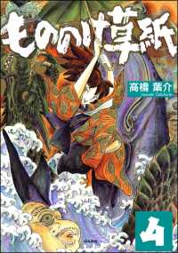
- 電子書籍
- もののけ草紙(分冊版) 【第4話】
-

- 電子書籍
- 白鯨 (上) 角川文庫


